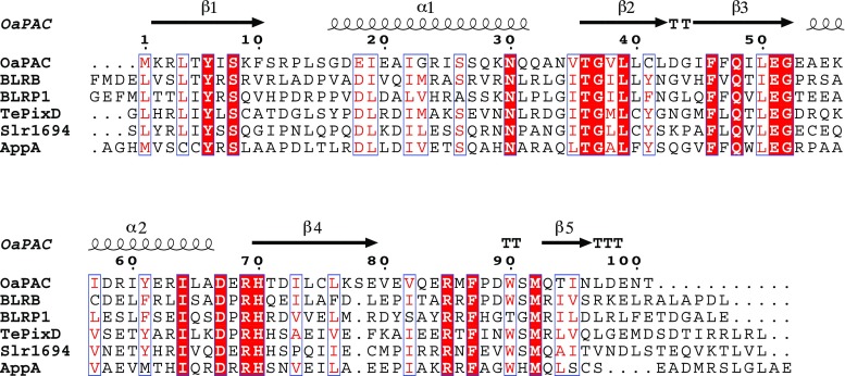
Fig. 1.
A sequence alignment of the six BLUF (Blue Light Using Flavin) domains for which independent experimental models have been deposited in the Protein Data Bank. The conserved residues, shown on a red background, are clustered around the flavin chromophore. Secondary structure elements from the crystal structure of OaPAC, a photo-activated adenyl cyclase (PAC) from Oscillatoria acuminata, are indicated with coils and arrows to indicate α-helices and β-strands, respectively, and turns are indicated by T. Tyr 8, Gln 48, Met 92 and Trp 90 of OaPAC form the quartet of residues that attract the most interest in functional studies. Asn 30 forms hydrogen bonds directly with the flavin. This figure was created with the program Espript (Robert and Gouet 2014)

