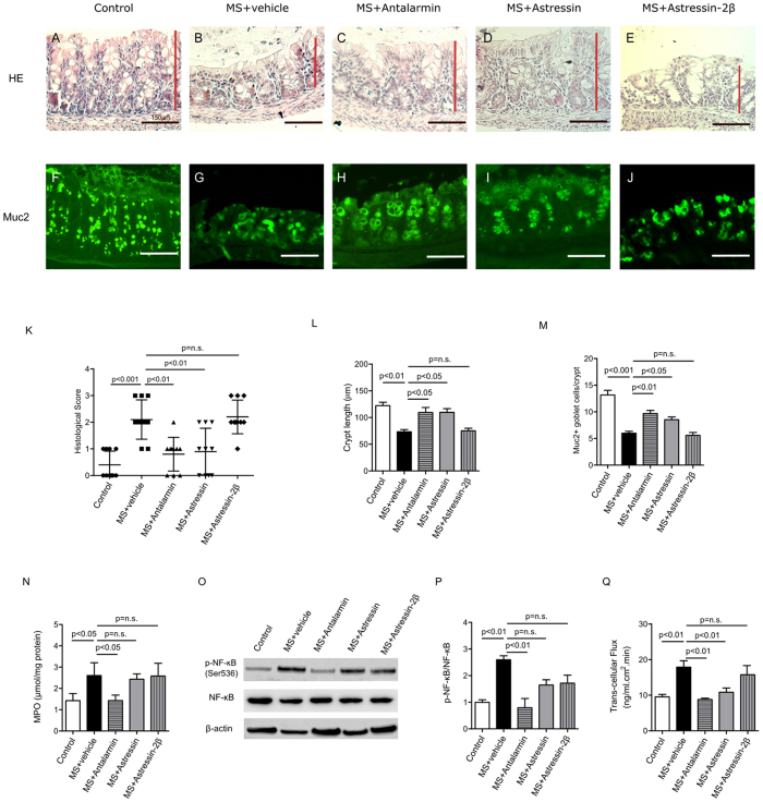
Figure 2. MS-induced intestinal epithelium injury was CRHR1 dependent.
Photomicrographs of hematoxylin and eosin (H&E) stained (A–E) and immunofluorescence of Mucin 2 (Muc2; mucous-forming protein) (F–J) in proximal colon in all experimental groups. Histological scores (K) were highest in MS, demonstrated injury in MS compared to control. Treatment with Antalarmin and Astressin prevented this MS-induced colonic injury, but not by Astressin-2β. Crypt length in μm (L) (red lines in photomicrographs A–E) and the number of Muc2+ goblet cells per crypt (M) were reduced by MS compared to control, and restored to control levels following Antalarmin and Astressin treatment. Astressin-2β did not prevent these MS-induced effects. Myeloperoxidase (MPO; μmol/mg protein) expression was increased in MS group and was reduced to a level similar to control by treatment with Antalarmin but not by treatment with Astressin or Astressin-2β (N). Western blot analysis of NF-κB showed an increase in the phosphorylated expression of NF-κB in MS, which was prevented by Antalarmin administration, but not by Astressin or Astressin-2β (O,P). Trans-cellular flux of HRP (ng/ml.cm2.min; Q) measured by Ussing Chamber was increased in MS and MS + Astressin-2β groups, compared to control, but not in MS + Antalarmin and MS + Astressin groups (P). Results are means, ±SD. p < 0.05 was considered significant.

