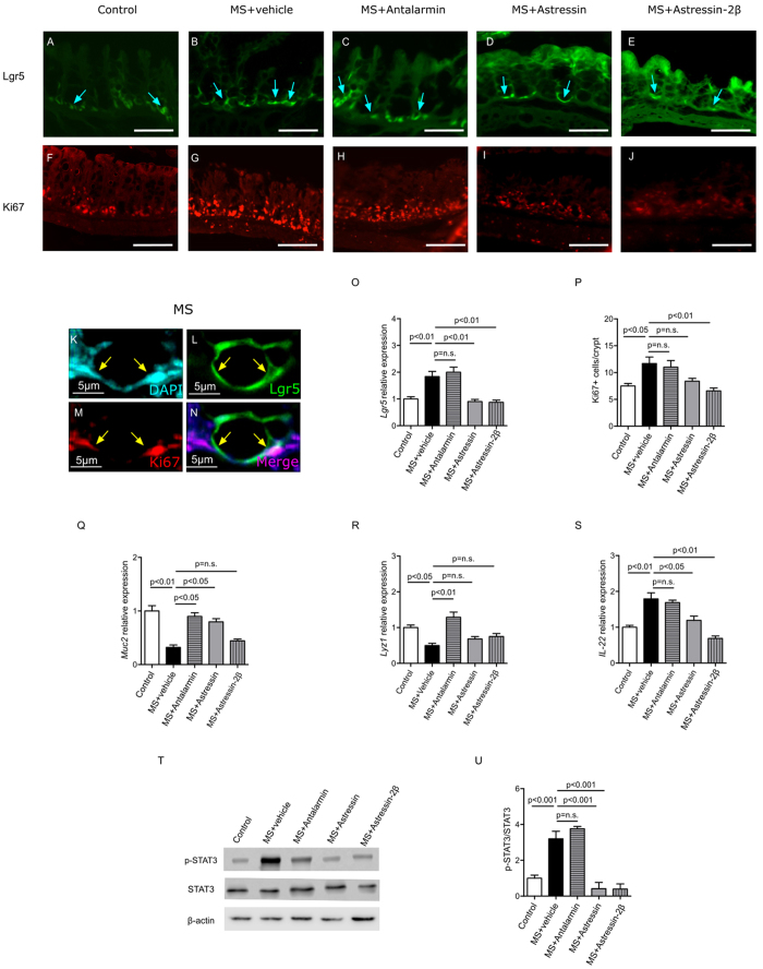
Figure 3. CRHR2 modulates the MS-induced increase in Lgr5+ intestinal epithelial stem cells (IESCs).
Fluorescent micrographs of intestinal stem cell marker Lgr5 (A–E, blue arrows) and cell proliferation marker Ki67 (F–J) in colonic tissues of the experimental groups. Lgr5 positive cells expressing Ki67 are shown in higher magnification (K–N, yellow arrows). Co-localization of Lgr5 and Ki67 markers in the intestine of pups subjected to MS suggested that Lgr5+ intestinal stem cells are proliferative. Relative gene expressions of Lgr5 (O) and the number of Ki67+ proliferating cells per crypt (P) in the colon were significantly increased by MS compared to control, and this increase was observed when Antalarmin was administered. In contrast, this increase was prevented by pre-treatment with both Astressin and Astressin-2β. Relative gene expressions of epithelial differentiation marker Muc2 (Q) and Lyz1 (R) are shown, The MS-induced decrease in Muc2 and Lyz1 expression was rescued by Antalarmin and Astressin, but not Astressin-2β. Relative gene expression IL-22 (S) and western blot of phosphorylated STAT3 (U) are shown. IL-22 and phosphorylated STAT3 increased in the MS group compared to the control; however, these elevations were both inhibited by Astressin and Astressin-2β, indicating the important role of CRHR2 in tissue repair in response to MS-induced injury. Results are means, ±SD. p < 0.05 was considered significant.

