Fig. 2.
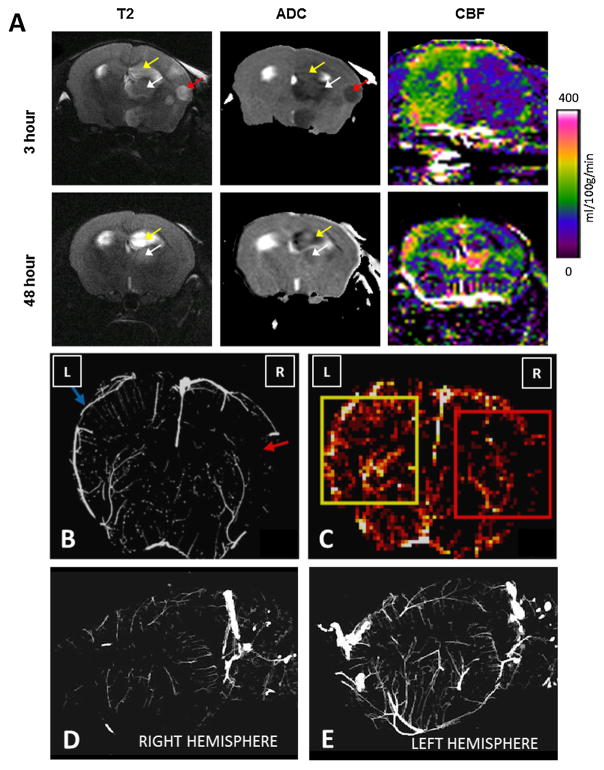
Cerebral perfusion and architecture are disrupted after injection of collagen in the mouse. Focal cerebral ischemia was induced by injecting six 1-μg boluses of collagen into the distal internal carotid artery of the mouse. (A) An 11.7 T magnetic resonance scanner was used to acquire T2-weighted (T2), apparent diffusion coefficient (ADC), and perfusion images on live mice 3 and 48 h after collagen injection. Regions with hyperintense T2 signals and hypointense ADC values were detected in the ipsilateral cortex (red arrow), hippocampus (yellow arrow), and thalamus (white arrow) at 3 h, accompanied by marked reductions in regional cerebral blood flow (CBF). At 48 h, T2 signals, ADC hypointensities, and CBF were improved, but abnormalities remained. (B) Micro-CT was performed on brains excised from mice 3 h after collagen injection. A coronal brain section shows disruption of vascular network in the middle cerebral artery territory and reduced vascular density ipsilateral to collagen injection (red arrow) compared to that in the contralateral hemisphere (blue arrow). (C) Micro-CT was used to construct cerebral heat maps that display average vessel density in the ischemic (red square) and contralateral brain (yellow square). Lighter colors represent higher vessel densities. (D) Right sagittal view from a micro-CT scan of the neurovasculature shows a poorly defined vascular network ipsilateral to collagen injection. (E) Left sagittal view of the neurovasculature contralateral to collagen injection. Images are representative of three separate experiments. (For interpretation of the references to color in this figure legend, the reader is referred to the web version of this article.)

