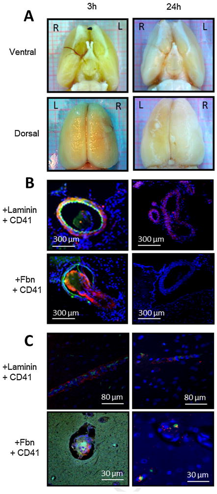
Fig. 7.
Injection of collagen into the middle cerebral artery (MCA) causes formation of platelet- and fibrin-rich thrombi in cerebral macro- and microvessels. Focal cerebral ischemia was induced by administering six 10-μl boluses of collagen near the right MCA orifice of the rat. At 3 and 24 h after collagen injection, brains were excised for histologic and immunofluorescent examinations of cerebral vasculature. (A) Ventral (upper panel) and dorsal (lower panel) gross images show thrombus in the MCA and its branches at 3 h (left panel) but not at 24 h (right panel) after collagen injection. (B) Immunofluorescent images show platelet- and fibrin-rich thrombi in cross sections of the MCA at 3 h (left panel) but not 24 h (right panel) after collagen injection. Upper panels are labeled with anti-laminin antibody (red), anti-CD41 platelet antibody (green), and anti-nuclear stain (blue). Lower panels are labeled with anti-fibrinogen/fibrin antibody (Fbn; red), anti-CD41 platelet antibody (green), and anti-nuclear stain (blue). (C) Coronal brain slices display platelet and fibrin deposition in cortical cerebral microvessels at 3 h (left panel) and 24 h (right panel) after collagen injection. Upper panels are labeled with anti-laminin antibody (red), anti-CD41 platelet antibody (green), and anti-nuclear stain (blue). Lower panels are labeled with anti-fibrinogen/fibrin antibody (red), anti-CD41 platelet antibody (green), and anti-nuclear stain (blue). Images are representative of four separate experiments. (For interpretation of the references to color in this figure legend, the reader is referred to the web version of this article.)

