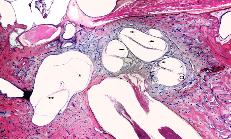
FIG. 1.

This is a 66-year-old white male with a clinical history of heart attack, occupational noise exposure, and left sensorineural hearing loss. He was treated with left cochlear implant (CI). Histopathology evaluation of the left ear showed: Simple mastoidectomy cavity, profound hydrops (arrows) in all turns of cochlea, saccular (*) and utricular hydrops (**), atrophy of stria vascularis (open arrow head) in lower basal turn, and hyalinization and fibrosis around electrode insertion site (CI).
