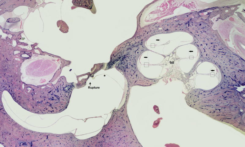
FIG. 4.

This is an 86-year-old male with a clinical history of Meniere’s disease and a Tack operation. Histopathology of the ear showed Tack hole seen in stapes footplate, severe endolymphatic hydrops, saccule hydropic and ruptured, severe loss ganglion cells, strial atrophy, and organ of Corti atrophy.
