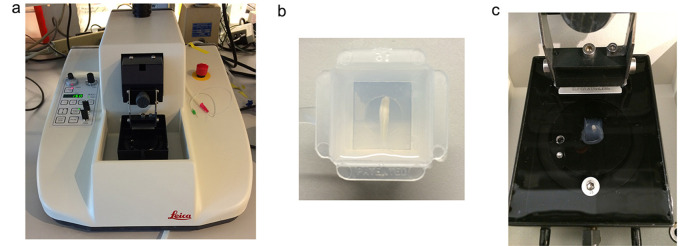
Figure 2. The experimental setup for sectioning the spinal cord.
a. Vibratome; b. A spinal cord segment placed horizontally in the agarose mold; c. The agarose cube containing the spinal cord is removed from the mold, rotated 90° and glued to the vibratome stage in the proper orientation for coronal sections.

