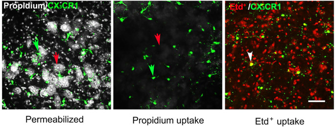
Figure 3. Uptake of propidium and Etd+ by glial cells and neurons in the ventral horn of spinal cord.
(Left panel) Slices were fixed overnight with 4% PFA and permeabilized, and then incubated in 5 μM PropI2 for 10 min and mounted. Motor neurons (red arrow) and microglia (green arrow) were identified by the morphology and by expression of EGFP, respectively, in acute slices prepared from CX3CR1EGFP/+ mice. In these slices, CX3CR1 and EGFP expressing cells are brain resident macrophages, a population mostly comprised of microglia. (Middle and right panels) In separate experiments, slices were maintained in ACSF saturated with 95% O2/5% CO2 for 1 h after sectioning and incubated with 5 μM PropI2 or 10 μM EtdBr for 10 min without permeabilization, rinsed and then fixed in 4% PFA before mounting. Under these conditions, there was little Prop2+ uptake in neurons (red arrow) and microglia (green arrow, middle panel). In contrast, Etd+ uptake was observed in CX3CR1EGFP/+ microglia (white arrow, Etd+ uptake plus EGFP expression), although it was rare in motor neurons (identifiable as round dark areas without small red cells, right panel, compare to left panel). The small red cells between EGFP- Etd+ cells and surrounding motoneurons are astrocytes, as was shown by GFAP immunolabeling (see Garré et al., 2016 ). Scale bar = 50 μm.

