Fig. 3.
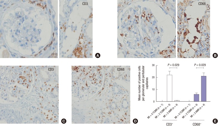
Intraluminal cell types in glomeruli and peritubular capillaries by immunohistochemistry. (A-D) Representative figures for CD3+ T cells (A) and CD68+ macrophages (B) in AMR with MI ≥ 2 and CD3+ T cells (C) and CD68+ macrophages (D) in TCMR with MI ≥ 2 (immunoperoxidase, original magnification × 200). (E) Comparisons of mean numbers of positive cells for CD3 and CD68 in glomerular and peritubular capillaries in biopsies with AMR with MI ≥ 2 (n = 4) versus TCMR with MI ≥ 2 (n = 4) versus TCMR with MI = 0 (n = 1). The average numbers of CD3+ T cells and CD68+ macrophages were significantly increased in TCMR with MI ≥ 2 biopsies (P = 0.029) and AMR with MI ≥ 2 biopsies (P = 0.029), respectively.
AMR = antibody-mediated rejection, MI = microcirculation inflammation, TCMR = T cell mediated rejection.

