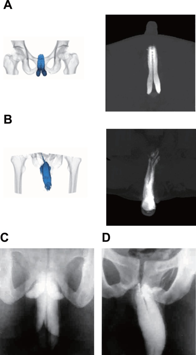
Figure 1. Image of corpora cavernosum detected by the cavernosography with 320-row DVCT compared with an image taken by conventional cavernosography.

(A) Cavernosography with 320-row DVCT image of regular cavernosa by; (B) cavernosography with 320-row DVCT image of cavernosal venous leakage by; (C) conventional cavernosonography image taken of the regular cavernosa by; (D) conventional cavernosa image of the cavernosal venous leakage by DVCT.
