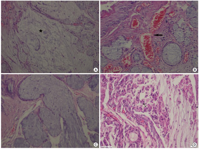
Fig. 5.

(A) Mucin pools in wide areas (star) due to the ruptured gland in the oropharyngeal tissue of the control group that received methotrexate (MTXG) (H&E, ×100). (B) Proliferated, dilated, and congested blood vessels (arrow) in the oropharyngeal tissue of the MTXG group (H&E, ×100). (C) Dilated ductal structures in the oropharyngeal tissue of the MTXG group (H&E, ×100). (D) Inflamed area (arrow) containing significant polymorphonuclear leukocytes in the oropharyngeal tissue of MTXG group (H&E, ×40).
