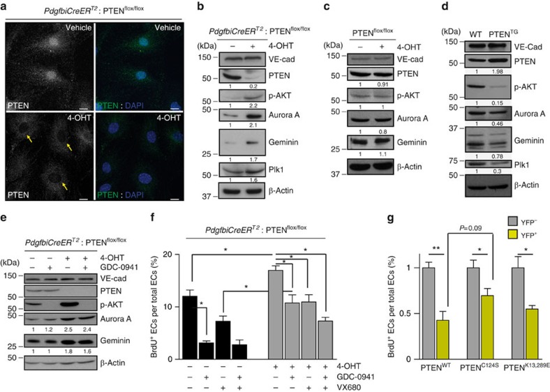
Figure 5. Catalytic and non-catalytic roles of PTEN regulate EC proliferation.
(a) Confocal images of PTEN (green) and DAPI immunofluorescence in PdgfbiCreERT2; PTENflox/flox mECs treated with vehicle or with 4-OHT for 96 h. Yellow arrows indicate the lack of PTEN staining in the nucleus of PTEN null cells. Scale bars, 10 μm (n=3). (b–d) Exponentially growing mECs were lysated, followed by immunoblotting using the indicated antibodies. PdgfbiCreERT2; PTENflox/flox (b) and PTENflox/flox (c) mECs were treated for 96 h with vehicle or 4-OHT. (d) WT and PTENTG mECs were cultured for 48 h before cell lysis and immunoblotting. (e) PdgfbiCreERT2; PTENflox/flox mECs were treated for 96 h with vehicle or 4-OHT. Before cell lysis, cells were pretreated for 2 h with GDC-0941 (1 μM). The quantification of the relative immunoreactivity of each protein normalized to β-actin is represented as the mean from at least three different experiments in b–e. Molecular weight marker (kDa) is indicated. (f) Exponentially growing control and PTENiΔEC/iΔEC mECs were treated for 48 h with test compounds or vehicle, and then were pulsed with BrdU for 2 h and subjected to immunostaining analysis. Inhibitors and doses used were as follows: GDC-0941 (pan-class I PI3K inhibitor; 1 μM) and VX680 (Aurora Kinase inhibitor; 0.5 μM). Data shown are means of four independent experiments. (g) PdgfbiCreERT2; PTENflox/flox mECs were infected with PTENWT, PTENC124S or PTENK13,289E, treated with 4-OHT for 72 h, plated for 48 h in the presence of doxycycline, pulsed with BrdU for 2 h and subjected to immunostaining analysis. Data shown are the means of six independent experiments. Error bars are s.e.m. *P<0.05 and **P<0.01 were considered statistically significant. Statistical analysis was performed by nonparametric Mann–Whitney test.

