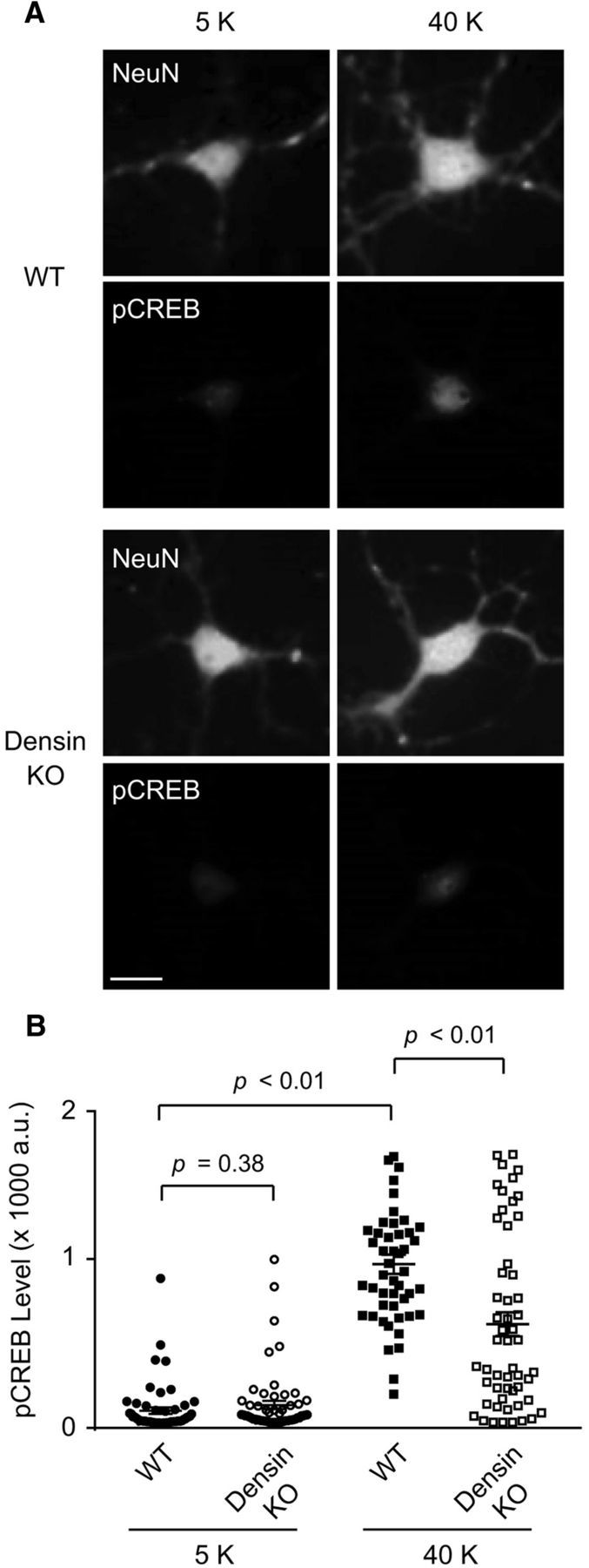
Figure 8.

Excitation–transcription coupling is reduced in densin KO neurons. A, Epifluorescence images of cortical neurons from WT and densin KO mice exposed to 5 or 40 mm K+ for 3 min and processing for double-label immunofluorescence with antibodies against pCREB and the neuronal marker, NeuN. Images are representative of three independent experiments. B, Quantification of the intensity of pCREB fluorescence (arbitrary units, a.u.) in WT and densin KO cortical neurons (n = 50–58 neurons per condition from three different sets of cultures). Statistical significance was determined by Student's t test.
