Figure 5.
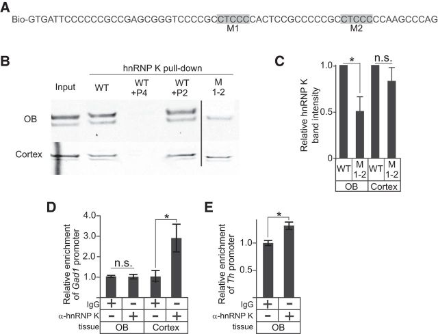
Indirect regulation of Gad1 promoter activity by hnRNP K. A, Biotinylated oligonucleotide sequence of the WT Gad1 promoter C-rich region with CT elements that were mutated highlighted in gray (M1 and M2). B, Western blots for hnRNP K from protein pull-down assays using WT and mutant (M1–2) oligonucleotides with either mouse OB or cortical nuclear lysate. Vertical line indicates the removal of intervening lanes. C, Relative Western blot band intensities for hnRNP K pulled down with either the WT or M1–2 oligonucleotides in B. D, ChIP assays for hnRNP K show occupancy on the Gad1 promoter in cortical, but not OB, tissue. E, ChIP assays for hnRNP K show occupancy on the Th promoter in OB tissue. *p < 0.01. Together, these findings indicate that hnRNP K is not recruited to the Gad1 promoter in the OB in vivo, and only indirectly associated with Gad1 promoter in the cortex.

