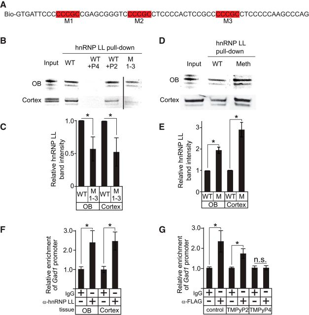
Figure 7.
hnRNP LL interacts with the Gad1 promoter C-rich strand. A, WT sequences of the biotinylated oligonucleotide used for pulldown of hnRNP LL. Red boxes indicate CG elements that were mutated. B, Western blots for hnRNP LL from protein pull-down assays using WT and mutant (M1–3) oligonucleotides with either mouse OB or cortical nuclear lysate. Vertical line indicates the removal of intervening lanes. C, Relative Western blot band intensities for hnRNP LL pulled down with either the WT or mutant (M1–3) oligonucleotides. D, Western blots for hnRNP LL from protein pull-down assays using WT and methylated oligonucleotides with either mouse OB or cortical nuclear lysate. The methylated oligonucleotide sequence was identical to the WT, except that all cytosine nucleotides in CpG motifs were methylated. E, Relative Western blot band intensities for hnRNP LL pulled down with either the WT or methylated oligonucleotides. F, ChIP assays for hnRNP LL show occupancy on the Gad1 promoter in both cortical and OB tissue. G, ChIP assays show that hnRNP LL occupancy on the Gad1 promoter in NT2 cells is blocked when TMPyP4, but not TMPyP2, is present. *p < 0.01.

