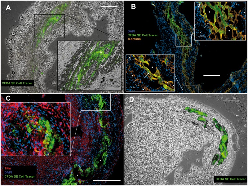
Fig 2. Genetically purified iPSC-derived CMs form mature grafts in vivo.
A+B: CFDA SE cell tracer positive iPSC-CM grafts 7 days after intramyocardial transplantation: Adjacent to the host myocardium iPSC-CMs align in a parallel, longitudinal fashion and exhibit sarcomeric structures (arrows). Within central portions of broader grafts (approximately > 200 μm) they maintain a small, round shape (* in A). In the infarct penumbra iPSC-CMs lie in close proximity to host CMs (arrowheads in B1), occasionally with direct cell contact (arrowheads in the bottom right corner of B1). Inside the infarct area iPSC-CMs are typically surrounded by infiltrating host cells (arrowheads in B2). Tissue disruption during histological preparation (* in B1+2) indicates loose cell adhesion within iPSC-CM grafts. C+D: CFDA SE cell tracer positive iPSC-CM graft 28 days after intramyocardial transplantation: The cell tracer remains visible 28 days after engraftment, but iPSC-CMs develop an amorphic appearance. Sarcomeric structures are not observed. Vacuoles form during histological preparation (* in C). A+D: brightfield overlay. Scale bars: 400μm.

