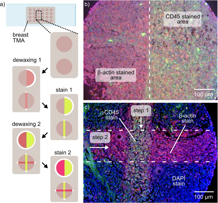
Fig 3. Multiplexed fluorescence-based IHC using tissue lithography.
(a) Schematic of the multiple dewaxing and staining steps performed. (b) Overlay image of two fluorescence channels showing multiplexed immunostaining of β-actin (red channel) and CD45 (green channel). (c) Overlay image of three fluorescence channels showing microscale partial staining of a TMA core. The tissue microarray was mounted using DAPI (blue channel) to highlight the non-stained areas of the sample.

