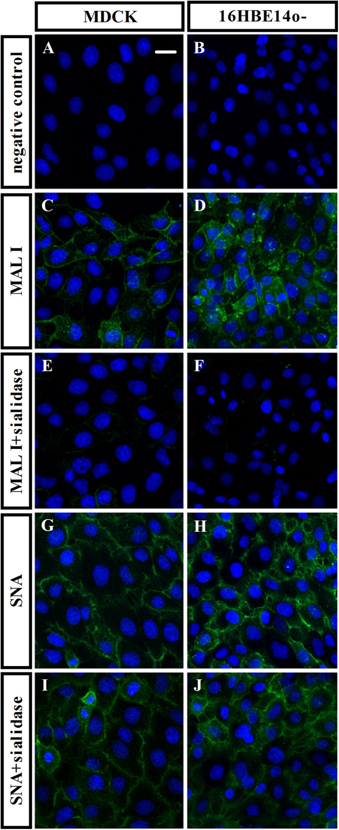
Fig 2. α-2,3 sialic acid receptor is expressed in 16HBE14o- cells.

Representative confocal images (800×) of 16HBE14o- cells (B, D, F, H and J) or MDCK cells (A, C, E, G and I) treated with DAPI (blue), to stain for nuclei, together with fluorescein-coupled antibody directed against α-2,3 sialic acid receptors, (green; C, D, E and F) or fluorescein-coupled antibody directed against α-2,6 sialic acid receptors, (green; G, H, I and J) or without fluorescein-lectin staining (negative control; A and B). The effect of sialidase on sialic acid receptors expression was performed by incubating cells with 1 U/ml sialidase 1 hr before stained with MAL I (MAL I+sialidase; E and F) or SNA (SNA+sialidase; I and J). Scale bar is 20 μm.
