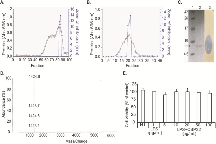
Fig 2.
(A) Gel filtration chromatography on a Sepharose CL-6B column (2.2 cm × 116 cm). The proteins were eluted at a flow rate of 5 mL/min. (B) Gel filtration chromatography on a Sephadex G-50 column (1.5 cm × 70 cm). The proteins were eluted at a flow rate of 1 mL/min. Determination of the molecular weight. (C) Tricine SDS-PAGE and activity staining of CSP32. Tricine SDS-PAGE: Lane 1, protein molecular weight markers with the corresponding value in kDa on the left; Lane 2, purified CSP32; Lane 3, activity staining (in situ). (D) MALDI-TOF-MS. (E) Cell viability was measured after 24-h incubation. Survival rates were tested by an MTT assay in RAW 264.7 cells. The cells were incubated in the presence or absence of 5–100 μg/mL CSP32 for 24 h. Each bar shows mean ± SD of three independent experiments performed in triplicate.

