
Figure 3.
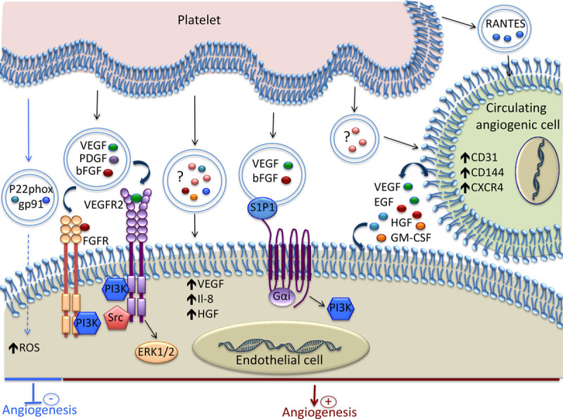
Mechanisms involved in the modulation of Angiogenesis by platelet-derived extracellular vesicles (EVs). Platelet-derived EVs contain various growth factors and chemokines that induce proangiogenic signaling in endothelial cell (EC). Spingosine-1-phosphate (S1P1), present on the EV surface, induces PI3K activation and, together with VEGF and bFGF, promotes angiogenesis. The EVs released by platelets stimulate the proangiogenic potential of circulating angiogenic cells by increasing their expression of both membrane molecules and soluble factors. Platelet-derived EVs can inhibit angiogenesis by transferring the p22phox and gp91 subunits of NADPH oxidase and increasing the oxidative stress in EC. ? indicates that the exact content of the EVs is not reported; bFGF, basic fibroblast growth factor; EGF, epidermal growth factor; GM-CSF, granulocyte-macrophage colony-stimulating factor; HGF, hepatocyte growth factor; NADPH, nicotinamide adenine dinucleotide phosphate; PI3K, phosphoinositide 3-kinase; RANTES, regulated on activation, normal T-cell–expressed and secreted; ROS, reactive oxygen species; VEGF, vascular endothelial growth factor; and VEGFR, vascular endothelial growth factor receptor.