
FIGURE 7.
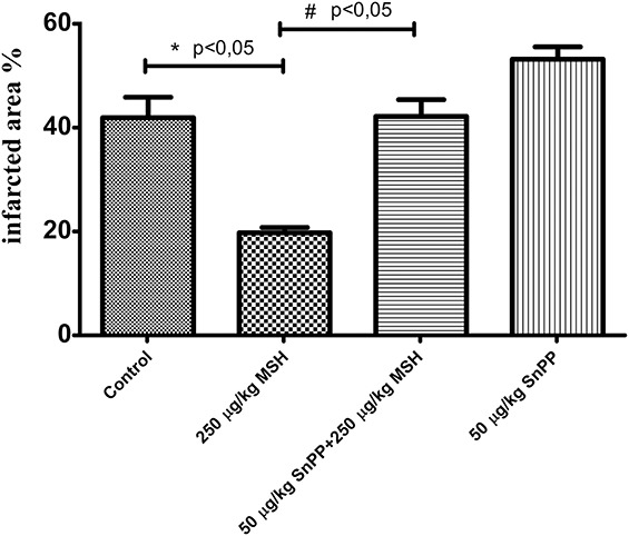
Effect of α-MSH and HO-1 inhibition on infarcted zone magnitude (protocol II). Rats administered subcutaneous saline (group II-a), or subcutaneous 250 μg/kg body weight α-MSH (group II-b), or intraperitoneal injections of 50 μg/kg body weight SnPP plus 250 μg/kg body weight subcutaneous α-MSH (group II-c), or 50 μg/body weight SnPP (group II-d), followed by sacrifice and ischemia/reperfusion injury during isolated working hearts according to protocol II. Staining of infarcted tissue was accomplished by administration to working hearts of 1% TTC solution in phosphate buffer, through the side arm of an aortic cannula. Hearts were sliced transversely, blotted dry, placed in between microscope slides, and scanned. Infarct areas of each slice were traced, and the respective areas were calculated by pixel density analysis. *P < 0.05 for comparison of infarcted areas to control. #P < 0.05 for comparison of infarcted areas to α-MSH–pretreated (250 μg/kg) group.