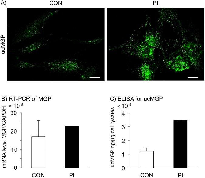
Fig 4. Increased level of ucMGP in GGCX dermal fibroblasts.
(A) Immunofluorescence staining with an antibody specific for ucMGP. The level of ucMGP was higher in GGCX dermal fibroblasts (Pt) than in normal dermal fibroblasts (CON). The bar depicts 50 μm (original magnification, ×400). (B) RT-PCR analysis of MGP mRNA levels in normal (CON) and GGCX (Pt) dermal fibroblasts. The MGP mRNA levels in normal dermal fibroblasts are shown as mean ± SD (n = 4) and those in GGCX dermal fibroblasts as mean. (C) Quantification of ucMGP in normal (CON) and GGCX (Pt) dermal fibroblasts. Values in normal dermal fibroblasts are shown as mean ± SD (n = 3) and those in GGCX dermal fibroblasts as mean. These experiments were performed at least twice, and a representative data set is shown.

