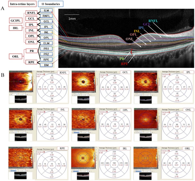
Fig 2. Representation of intra-retinal layer segmentation and thickness measurement.
(A): Segmentation of the retinal boundaries in a normal eye by Spectralis SD-OCT: 1 = inner limiting membrane (ILM); 2 = retinal nerve fiber layer (RNFL); 3 = ganglion cell layer (GCL); 4 = inner plexiform layer (IPL); 5 = inner nuclear layer (INL); 6 = outer plexiform layer (OPL); 7 = outer limiting membrane (OLM); 8 = myoid zone of the photoreceptor layer (PR1); 9 = ellipsoid component of the photoreceptor layer (PR2); 10 = retinal pigment epithelium (RPE); and 11 = Bruch’s membrane. (B): Representative 9 intra-retinal layer measurements within a standard ETDRS grid: average volume and thickness measurements for the fovea subfield, inner and outer circle subfields and 9 sectors.

