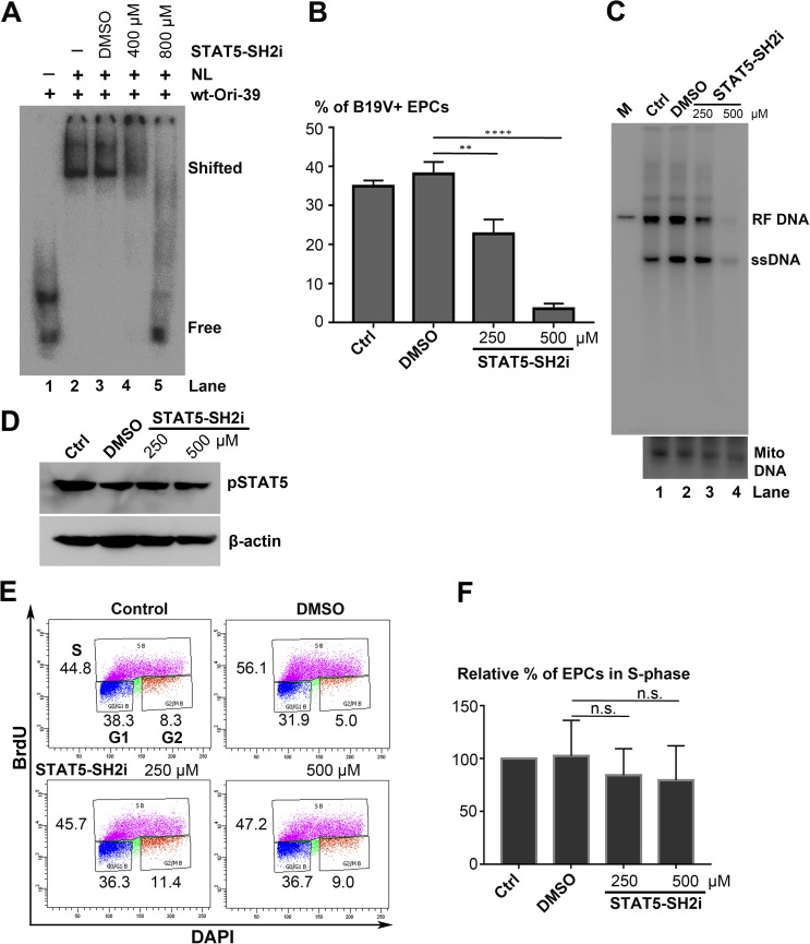
Fig 5. Blockage of interaction between STAT5 and B19V Ori DNA inhibits B19V replication.
(A) STAT5-SH2 inhibitor (STAT5-SH2i) abolishes the shift of viral Ori in EMSA. UT7/Epo-S1 nuclear lysate (NL) was incubated with 32P-labelled wt-Ori-39 probe (lanes 2–5) with the addition of DMSO (lane 3) or STAT5-SH2i at 0.4 mM (lane 4) and 0.8 mM (lane 5). (B-D) STAT5-SH2i significantly inhibits viral DNA replication. CD36+ EPCs were incubated with either DMSO or STAT5-SH2i (250 μM or 500 μM), 6 h prior to infection. At 48 h post-infection, cells were collected and subjected either to (B) flow cytometry analysis for the B19V-infected (B19V+) cell population, with an anti-capsid antibody, or to (C) Hirt DNA extraction for Southern blot analysis with a B19V M20 DNA probe (upper panel), with mitochondrial DNA (Mito DNA) probed as a loading control (lower panel), or to (D) protein extraction for Western blotting with anti-pSTAT5 and anti-β-actin (E&F) Effect of STAT5-SH2i on cell proliferation. CD36+ EPCs were treated with either DMSO or STAT5-SH2i (250 μM or 500 μM), and were then incubated with BrdU to perform BrdU incorporation assays. (E) Results of a representative cell-cycle analysis. (F) Relative fold changes in the S-phase cell population of each group shown with means and standard deviations of three independent experiments. P values are calculated using one-way ANOVA and Tukey-Kramer post-test (P>0.05), compared with DMSO control. **** denotes P<0.0001, ** P<0.01, and n.s. no statistical significance.

