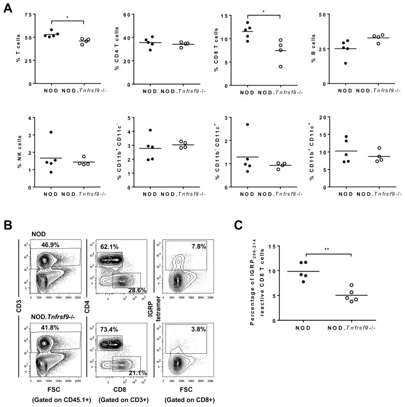
Figure 3. Intra-islet accumulation of CD8 T cells is proportionally reduced in the NOD.Tnfrsf9−/− strain compared to NOD mice.
(A) Flow cytometric analysis of various islet infiltrating leukocyte populations. Islets were isolated from 9–12 week old NOD and NOD.Tnfrsf9−/− female mice and dissociated into single cell suspension. Leukocytes were identified by CD45.1 expression and the frequencies of different cell populations, including total T cells (CD3+), CD4 T cells (CD3+ CD4+), CD8 T cells (CD3+ CD8+), B cells (CD19+), NK cells (CD3− DX5+), and myeloid cells (CD11b+ CD11c−, CD11b+ CD11c+, and CD11b− CD11c+) were calculated as percentages of total CD45.1+ cells. Each symbol represents cells pooled from 2–7 mice. *P < 0.05 by Mann-Whitney test. (B and C) The frequencies of islet associated IGRP206-214 reactive CD8 T cells in NOD and NOD.Tnfrsf9−/− female mice by MHC class I tetramer staining. Islet cells pooled from 2–5 mice (10–12 weeks old) were stained with antibodies against CD45.1, CD3, CD4, and CD8 as well as the IGRP206-214 MHC I tetramer. (B) One representative experiment is shown. (C) Summarized results from five independent experiments. **P < 0.01 by Mann-Whitney test.

