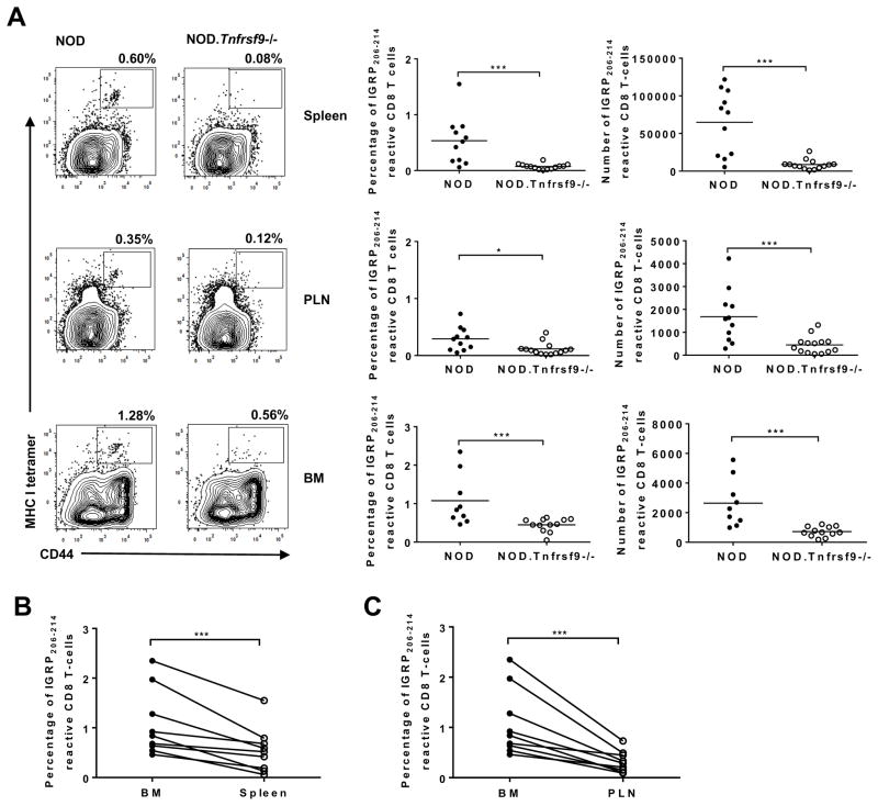
Figure 4. The frequencies and numbers of IGRP206-214 reactive CD8 T cells in the spleen, PLN, and BM are significantly lower in NOD.Tnfrsf9−/− than in NOD mice.
(A) Cells were isolated from the spleen, PLN, and BM, and strained with antibodies against CD8 and CD44 as well as IGRP206-214 loaded MHC class I tetramers. Representative flow cytometry profiles are shown in the left panels. The percentages and numbers of IGRP206-214 MHC class I tetramer staining of the spleen, PLN, and BM from 10–14 week old NOD and NOD.Tnfrsf9−/− female mice from 4 independent experiments are summarized respectively in the middle and right panels. In one experiment, BM from two mice of each strain were not analyzed. *P < 0.05, ***P < 0.005 by Mann-Whitney test. (B and C) The frequency of IGRP206-214 reactive CD8 T cells is higher in the BM than in the spleen (B) and PLN (C). Paired analysis of IGRP206-214 MHC class I tetramer staining for BM versus spleen and BM versus PLN in NOD mice. ***P < 0.005 by Wilcoxon matched-pairs signed rank test.

