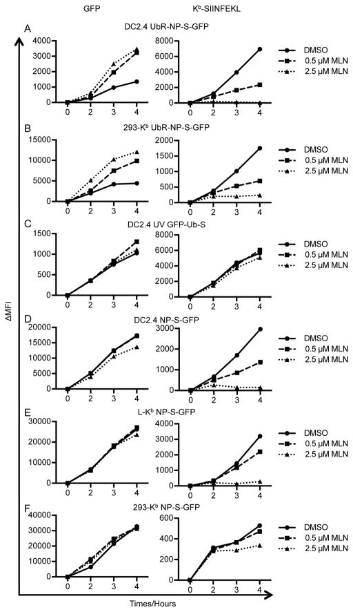
Figure 4. MLN7243 selectively inhibits Kb-SIINFEKL presentation from rVV-expressed proteins.
DC2.4 (A, C and D), L-Kb (E), and 293-Kb (B and F) cells were infected for 1 h with rVV expressing UbR-NP-S-GFP (A and B), partially UV-inactivated rVV expressing GFP-Ub-S (C), or rVV expressing NP-S-GFP (D–F). Cells were then cultured in the presence of DMSO, 0.5 μM MLN7243, or 2.5 μM MLN7243 and harvested at indicated time points. Levels of GFP (left panels) and surface Kb-SIINFEKL (right panels) were determined by flow cytometry. ΔMFI: medium fluorescent intensity after background subtraction.

