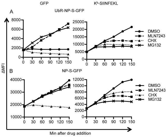
Figure 5. Fine kinetics of inhibitor blockade.
L-Kb cells were infected with rVVs expressing UbR-NP-S-GFP (A) or NP-S-GFP (B) for 1 h. At 3 h, DMSO, MLN7243, CHX or MG132 were added into cell cultures. Cells were harvested at indicated time points after drug addition. Levels of GFP (left panels) and surface Kb-SIINFEKL (right panels) were determined by flow cytometry. ΔMFI: medium fluorescent intensity after background subtraction.

