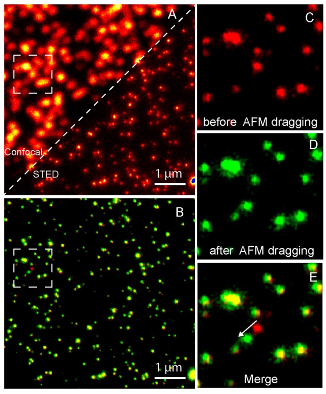
Figure 8.
Nanomanipulation of a 40 nm fluorescent bead using the correlative STED/AFM technique. (A) A comparison of confocal and STED images which shows that STED has a higher resolution. (B) Merged image of STED images acquired before (red) and after (green) AFM dragging of the same area. The overlay of both colors shows stationary beads in yellow. Magnified STED images acquired before (C) and after (D) AFM dragging, and the corresponding merged image (E), which clearly shows that the movement made by AFM at a subdiffraction distance. Reproduced from [64] with permission.

