Fig. 3.

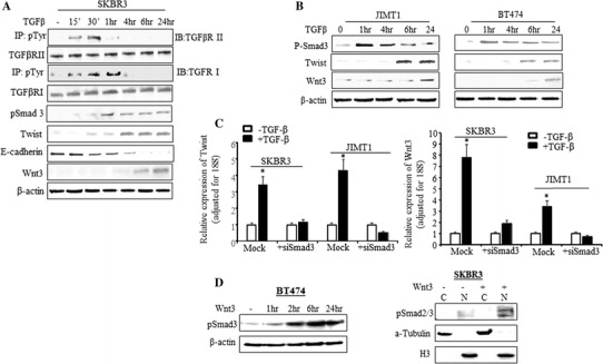
TGF-β-induced EMT and Wnt3 are Smad3 dependent. a SKBR3 cells were treated with TGF-β for 15 min to 24 h. Total protein was extracted at the indicated time points. To measure the phosphorylated TGF-βRI and TGF-βRII, immunoprecipitation was performed by incubating protein lysis with phosphor-Tyrosine antibody first followed by Western blot analysis with antibodies specific to TGFβRI and TGFβRII. The phospho-Smad3 (pSmad3), Twist, E-cadherin, and Wnt3 protein levels were also determined by Western blot analysis. The β-actin was used as loading control. b BT474 and JIMT1 cells were treated with TGF-β from 1 to 24 h and Western blot analysis was performed with the indicated antibodies. c Cells were treated with siSmad3 or negative sequence (Mock) for 48 h. TGF-β was added into the media containing siSmad3 or negative sequence after 24 h siSmad3 treatment. RT-qPCR was performed with Twist (left) and Wnt3 (right) primers. The bars indicate relative expression of the indicated genes adjusted to 18S (mean ± SD from three determinations), *p < 0.01 compared to untreated cells. d In the left panel, BT474 cells were treated with recombinant human Wnt3 protein at the indicated time and total protein was extracted. The pSmad3 was determined by Western blot analysis. The β-actin was used as loading control. In right panel, SKBR3 cells was treated with or without recombinant human Wnt3 protein for 24 h and then cytoplasmic and nuclear fragment was extracted. The pSmad2/3 was determined by Western blot analysis. The α-Tubulin and H3 were used for loading control of cytoplasm and nuclear protein respectively
