
FIGURE 2.
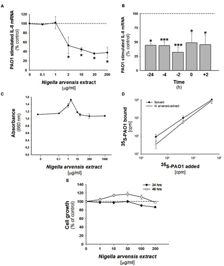
Effect of N. arvensis extracts in IB3-1 cells. (A) Effect of N. arvensis extracts on IL-8 mRNA expression in IB3-1 cells. IB3-1 cells were treated with the chloroform extract (solved in EtOH/DMSO 95/5) (A) at different concentrations (0.1–200 μg/ml) for 16 h before infection with PAO1 (100 CFU/cell) for further 4 h. IL-8 mRNA expression was quantified by qRT-PCR. Expression of IL-8 mRNA was measured by Real-Time qPCR and obtained by comparing the ratio IL-8 and the housekeeping gene GAPDH between non-infected and infected cells. The results are expressed as the % of untreated cells. Data are mean ±SEM of three independent experiments performed in duplicate. Dashed line corresponds to cells treated with solvent alone. (B) Effect of N. arvensis extract in PAO1 infected IB3-1 cells after different incubation times. The N. arvensis extract (10 μg/ml) was added to IB3-1 cells 24, 4, and 2 h before, simultaneously or 2 h post PAO1 infection (100 CFU/cell). IL-8 mRNA expression was measured as indicated in (A). Data are mean ±SEM of three independent experiments performed in duplicate. Dashed line corresponds to cells treated with solvent alone. (C) PAO1 growth. Bacteria were cultured overnight at 37°C in the presence of solvent or ranging doses (0.1–1000 μg/ml) of N. arvensis extract. Bacterial growth was monitored by absorbance measures at 660 nm. A representative experiment performed in duplicate is shown. (D) Adhesion of PAO1 to IB3.1 cells. 500,000 IB3-1 cells on Petri dishes, in duplicate, were treated for 24 h with 10 μg/ml N. arvensis extract. Different amounts of 35S-PAO1, expressed as CFU/well, were added to the wells and incubated as described in section “Materials and Methods”. Data reported in the figure are the specific binding calculated by subtracting counts obtained in the presence of 100-fold excess of non-labeled PAO1 and are expressed as CFU/well. Data are mean ±SEM of three independent experiments performed in duplicate. (E) Effects of N. arvensis extracts on cell growth in IB3-1 cells. IB3-1 cells were incubated with increasing concentrations (1–200 μg/ml) of the chloroform extract (solved in EtOH/DMSO) of N. arvensis for 24 and 48 h. Cell viability was measured by cytometer analysis. Data are expressed as % control (solvent) and are relative to a representative experiment performed in duplicate. Dashed line corresponds to cells treated with solvent alone.