
FIGURE 6.
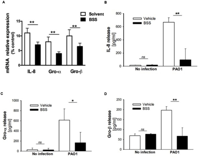
Effect of BSS on expression of neutrophil chemokines in CuFi-1 cells. (A) CuFi-1 cells were treated for 16 h with solvent alone or 100 nM BSS and then infected by PAO1 (10–50 CFU/cell) for 4 h. mRNA expression was measured as indicated in the legend of Figure 2. (B–D) Release of the neutrophil chemokines IL-8, Groα, and GROβ in the supernatants of CuFi-1 cells were measured by Bio-plex assay. CuFi-1 cells were treated as described for (A). Representative of at least three experiments performed in duplicate.