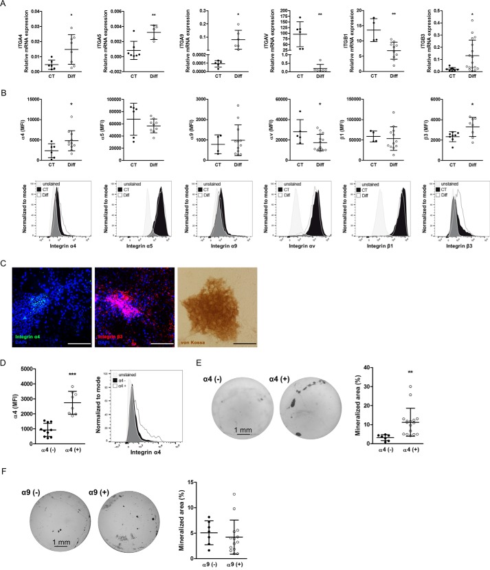
Figure 5.
Expression profile of integrin subunits relevant for EDA- and EDB-containing fibronectin on osteoblasts at baseline and after differentiation. A, mRNA expression of integrins α4, α5, α9, αv, β1, and β3 before and after differentiation. Primary newborn calvarial osteoblasts were cultured for 2–3 weeks in mineralizing medium and compared with cells cultured in medium without additives (biological replicates for α4: n = 6/8 in 2 experiments; α5: n = 7/5 in 1 experiment; α9: n = 5/6 in 2 experiments; αv: n = 6/6 in 1 experiment; β1: n = 4/10 in 3 experiments; and β3: n = 9/17 in 2 experiments). B, flow cytometry of osteoblasts stained for the mentioned integrin subunits showing the MFI in the first row and examples of the measurements in the second row (corrected to show equivalent numbers of cells), with the gray peaks representing the autofluorescence of the cells (biological replicates for α4: n = 6/11; α5: n = 6/9; α9: n = 4/12; αv: n = 5/15; β1: n = 4/12; β3: n = 8/8 in 3 experiments). C, mineralized nodules stained against α4 integrin (green), β3 integrin (red), and von Kossa confirm the presence of α4 within the nodules and β3 mostly within, but also around, the nodules. A sister well was stained against von Kossa to confirm mineralization. Osteoblasts mineralized for 2–3 weeks were stained against α4 and β3 and with von Kossa. Nuclei were stained with DAPI. Bars, 100 μm. D, osteoblasts were separated based on their α4 integrin subunit expression. Following isolation of α4-enriched and α4-depleted cells and culture for 7 days, flow cytometry was repeated and revealed persistent low expression of α4 in the depleted fraction and higher expression in the α4-enriched fraction (biological replicates: n = 10/8 in 4 experiments). E, α4-enriched osteoblasts (α4(+)) show enhanced differentiation compared with α4-depleted cells (α4(−)). Shown are examples of von Kossa staining and quantification of the stained area (biological replicates: n = 7/16 in 3 experiments). F, no differences in mineralization are found between osteoblasts that express α9 (α9(+)) and those that do not (α9(−)) (biological replicates: n = 7/14 in 2 experiments). Primary osteoblasts were stained for α4 or α9 and separated into two populations based on the expression of α4 or α9 and then differentiated for 2–3 weeks in mineralizing medium, stained by von Kossa, and evaluated. Results are expressed as mean ± S.D. (error bars). *, p < 0.05; **, p < 0.01; ***, p < 0.005.

