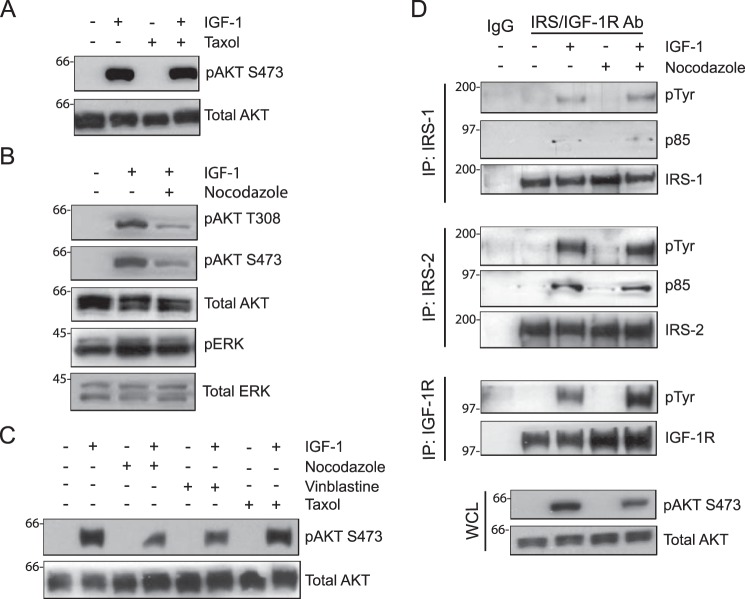
Figure 1.
Involvement of the microtubule cytoskeleton in IGF-1-stimulated AKT activation. MDA-MB-231 cells were treated with DMSO, Taxol (20 μm), nocodazole (1 μm), or vinblastine (20 nm) for 30 min and then stimulated with IGF-1 (20 ng/ml) for 5 min. A—C, aliquots of cell extracts containing equivalent amounts of total protein were immunoblotted with antibodies specific for Ser(P)-473AKT (pAKT S473), Thr(P)-308AKT (pAKT T308), or Thr(P)-202/Tyr-204ERK (pERK). The immunoblots were subsequently stripped and reprobed with total AKT and ERK-specific antibodies. D, aliquots of cell extracts containing equivalent amounts of total protein were immunoprecipitated (IP) with antibodies specific for IRS1, IRS2, or IGF-1Rβ subunit and immunoblotted with antibodies specific for phosphotyrosine (pTyr) and the p85 subunit of PI3K (p85). The Tyr(P) immunoblots were subsequently stripped and reprobed with IRS1, IRS2, or IGF-1Rβ-specific antibodies. Total cell extracts were also immunoblotted with antibodies specific for Ser(P)-473AKT and total AKT (whole-cell lysate (WCL), bottom panels). All immunoblots shown are representative of three independent experiments.

