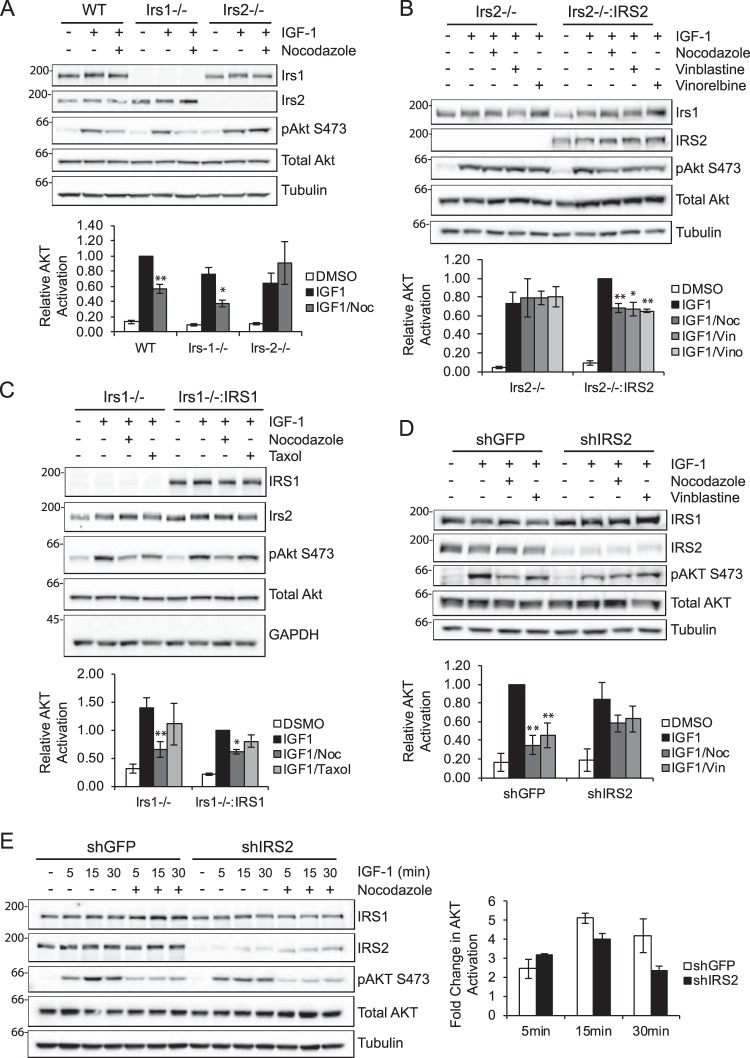
Figure 2.
Selective requirement of the microtubule cytoskeleton for IRS-2-mediated signaling. A, PyMT:WT, PyMT:Irs-1−/− and PyMT:Irs-2−/− cells were treated with DMSO or 20 μm nocodazole (Noc) for 1 h and then stimulated with IGF-1 (10 ng/ml) for 5 min. B, PyMT:Irs-2−/− cells transfected with empty vector (Irs2−/−) or IRS2 (Irs2−/−:IRS2) were treated with DMSO, 1 μm nocodazole, 20 nm vinblastine (Vin), or 20 nm vinorelbine (Vino) for 1 h and then stimulated with IGF-1 (10 ng/ml) for 15 min. C, PyMT:Irs-1−/− cells transfected with empty vector (Irs1−/−) or IRS1 (Irs1−/−:IRS1) were treated with DMSO, 1 μm nocodazole, or 10 μm Taxol for 1 h and then stimulated with IGF-1 (10 ng/ml) for 5 min. D, MDA-MB-231 cells expressing either an shRNA targeting GFP (shGFP) or IRS2 (shIRS2) were treated with DMSO, 1 μm nocodazole, or 20 nm vinblastine for 30 min and then stimulated with IGF-1 (10 ng/ml) for 30 min. E, MDA-MB-231 cells expressing either an shRNA targeting GFP or IRS2 were treated with DMSO or 1 μm nocodazole for 30 min and then stimulated with IGF-1 (10 ng/ml) for the time periods indicated. The data in the graph represent the -fold change in phospho-AKT between DMSO- and nocodazole-treated cells for each cell type. Aliquots of cell extracts containing equivalent amounts of total protein were immunoblotted with antibodies specific for IRS1, IRS2, Ser(P)-473AKT, total AKT, tubulin, or GAPDH. The data shown in the graphs for each immunoblot represent the mean ± S.E. of three independent experiments. *, p ≤ 0.05 relative to shGFP; **, p ≤ 0.01 relative to shGFP.

