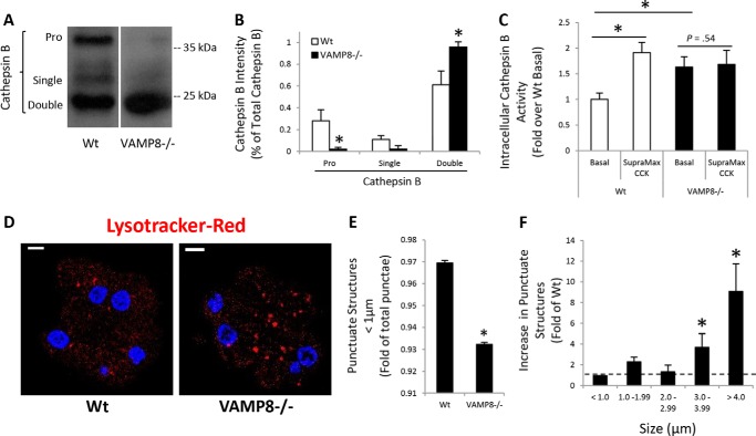
Figure 4.
VAMP8−/− have enhanced lysosomal CatB activity. CatB maturation was determined by immunoblotting (A) and quantified by densitometry (B) in WT or VAMP8−/− acinar lysates. Pro, proenzyme. C, isolated WT or VAMP8−/− acini were left untreated or stimulated with 10 nm CCK-8 for 60 min. Intracellular CatB activity was measured using a fluorogenic substrate. D, WT or VAMP8−/− acini were incubated with 50 nm Lysotracker Red DND-99 for 45 min and imaged by confocal microscopy (scale bars, 5 μm). E, Lysotracker Red punctate structures <1 μm were quantified by ImageJ. F, Lysotracker Red punctate structures binned to the indicated sizes for VAMP8 were quantified as -fold WT (data are the mean and S.E. *, p < 0.05). All experiments were generated from at least three separate acinar preparations, performed in duplicate.

