Figure 7.
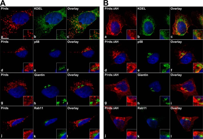
P/rdsΔAH forms inclusions that appear largely distinct from secretory pathway organelles in cultured cells. LSCM analysis of HEK AD293 cells stably expressing either WT P/rds (A) or mutant P/rdsΔAH (B) were double-labeled for indirect immunofluorescence with reagents directed against P/rds (red) and a marker (green) for ER (KDEL), ERGIC (p58), Golgi (giantin), or ERC (Rab11) organelles. Nuclei are counterstained with Hoechst 33342 (blue). Single optical sections from representative cells with moderate protein expression levels are shown. The majority of each protein was distributed in discrete puncta (WT) or large inclusions (P/rdsΔAH), which were distinct from the membranous organelles surveyed. No significant co-localization was observed between WT P/rds or P/rdsΔAH and any of the membranous compartments. Co-localization was quantified by Pearson's correlation coefficient (PCC) using the Nikon Elements software. For WT P/rds, the PCCs were as follows: KDEL, 0.61; p58, 0.47; giantin, 0.35; Rab11, 0.53. For mutant P/rdsΔAH, the PCCs were as follows: KDEL, 0.65; p58, 0.44; giantin, 0.40; Rab11, 0.42. 3D volume views reconstructed from Z-section stacks (including double labelings against KDEL and giantin) are provided as supplemental Figs. 5–8. Scale bar, 10 μm.

