Figure 8.
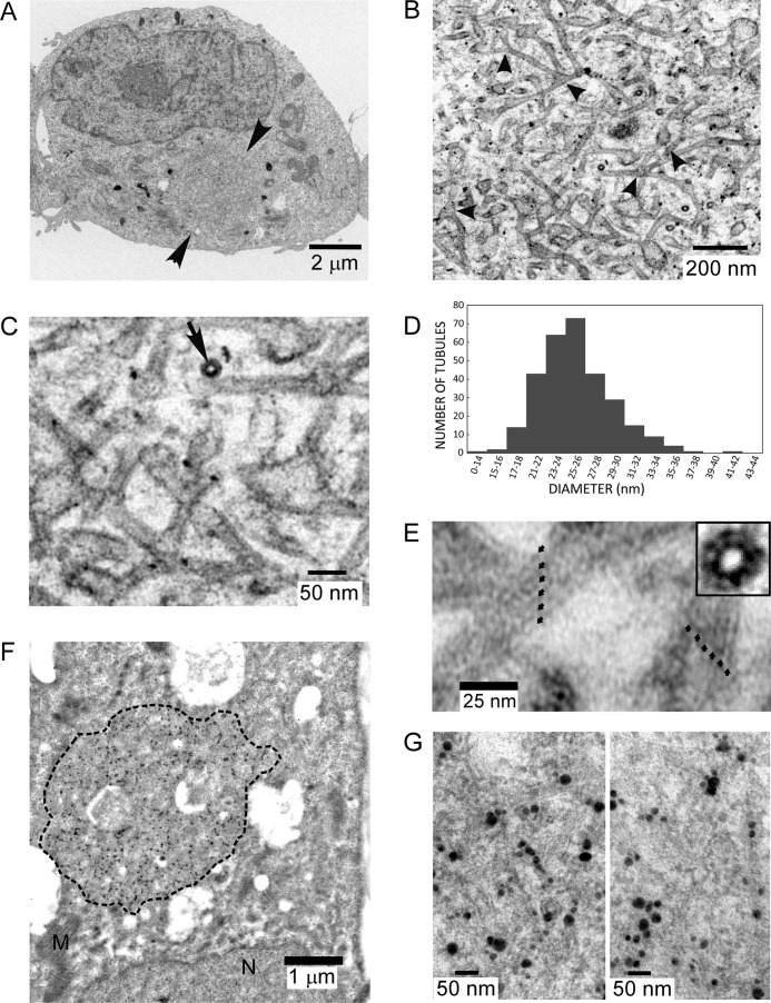
P/rdsΔAH displays robust membrane curvature-generating activity. A, transmission electron micrograph of a post-stained HEK AD293 cell stably expressing P/rdsΔAH shows a large inclusion (arrowheads), possessing dimensions similar to those documented by immunocytochemistry (Fig. 7B). B, higher magnification view of the inclusion, illustrating the dense network of tubulovesicular membranes within it; several three-way junctions are evident (arrowheads). C, higher magnification view from the same inclusion illustrates the trilaminar structure of the membranes, a high-curvature tubule sectioned in a transverse orientation (arrow), and several junctions. D, histogram illustrating diameters of tubules generated by P/rdsΔAH expression. E, tubules imaged at higher magnifications display regular substructure. Two tubules (∼26-27- nm diameters) showing striations along their longitudinal axes with a repeat of roughly 5.5 nm (small arrows). Inset, a transverse section shows a similar periodicity. F, transmission electron micrograph of a HEK AD293 cell stably expressing P/rdsΔAH, labeled with anti-P/rds monoclonal antibody Mab2B6 and anti-mouse-IgG nanogold followed by silver enhancement. The large inclusion (dotted perimeter) shows robust labeling. G, higher magnification views of immunogold inclusion labeling showing the presence of P/rdsΔAH in the tubulovesicular membranes. The reduced ultrastructural preservation (i.e. versus C) is typical for immunogold labeling experiments, because milder fixation conditions are needed to retain antigenicity.

