Figure 2.
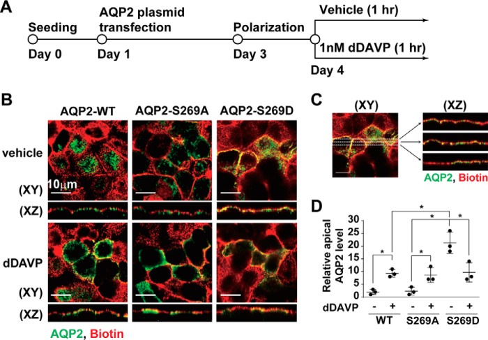
Phosphorylation-mimicking mutation at Ser-269 enhanced apical AQP2 retention in the mpkCCD cells. A, the experimental protocol. B, confocal immunofluorescence micrographs of the wild type (WT), S269A and S269D mutant AQP2 in mpkCCD cells stimulated with vehicle or dDAVP for 1 h. The apical plasma membrane was labeled with biotin. C, the image analysis protocol. Three lines were drawn across each X-Y plane image to produce three X-Z plane images. Relative apical AQP2 level was calculated as the AQP2 pixels that overlapped with the biotin pixels divided by the total AQP2 pixels. D, a scatter plot summarizing the imaging results. Values are mean ± S.D. Asterisk indicates significance p < 0.05, t test.

