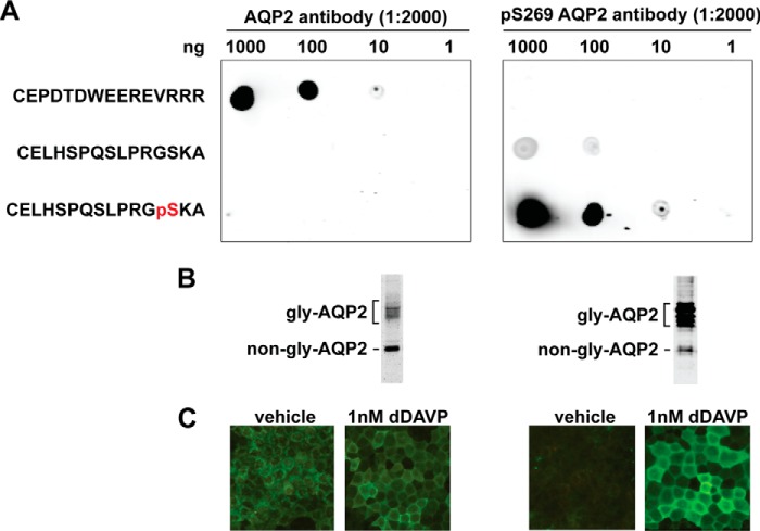
Figure 9.
AQP2 antibody characterization. A, dot blot tests for the specificity of the antibodies raised against the immunizing AQP2 peptide (CEPDTDWEEREVRRR) and the Ser-269-phosphorylated (pS269) AQP2 peptide (CELHSPQSLPRGpSKA). Serial diluted peptides were dotted on nitrocellulose membranes and incubated with the antibodies before detection. B, immunoblotting tests for the antibodies. Protein samples were from the mpkCCD cells exposed to dDAVP (1 nm) for 4 days. Gly-AQP2, glycosylated; non-gly-AQP2, non-glycosylated AQP2. C, immunofluorescence staining tests for the antibodies. The mpkCCD cells went through the experimental protocol shown in Fig. 1A.

