. 2017 Mar 14;16:265–277. doi: 10.17179/excli2017-119

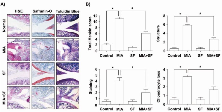
Figure 2. Histological evaluation of joints activity after administration with SF in MIA-induced OA rats. Rats were injected with 3 mg MIA in the right knee. SF was administered orally daily for 3 weeks after MIA injection. (A) The knee joints of OA rats treated with either SF or vehicle control were stained with H&E, Safranin O and toluidine blue. (B) The joint lesions were graded on a scale of 0-13 using the modified Mankin scoring system, giving a combined score for cartilage structure, cellular abnormalities, and matrix staining. Data were expressed as mean ± SD (n = 8) (*p < 0.05 MIA group vs. normal control group; #p < 0.05 SF+MIA treatment groups vs. MIA group).

Figure 2