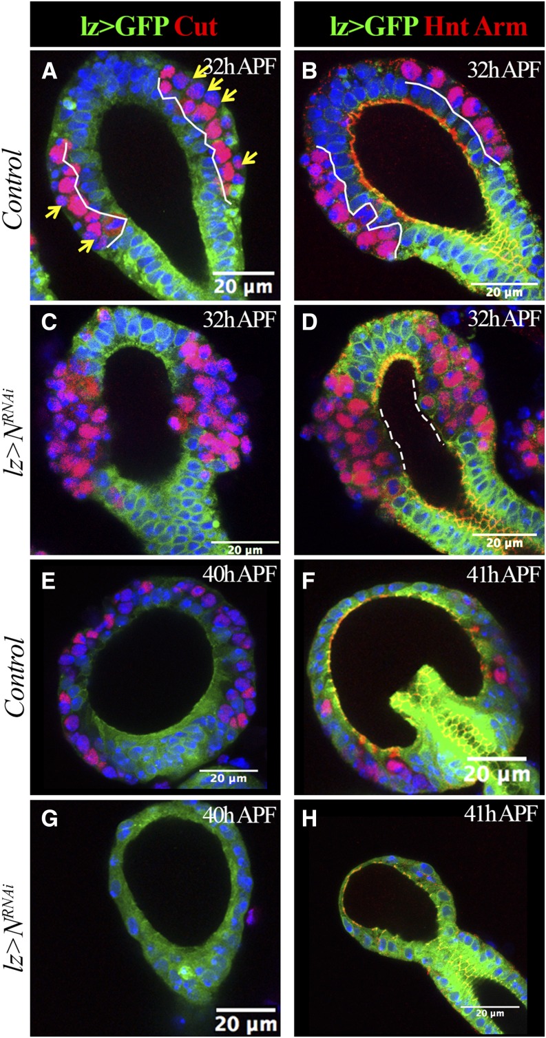
Figure 3.
Loss of LEP causes dissociation of secretory units from the central lumen. lz-Gal4 driving expression of UAS-mCD8:GFP (lz > GFP) is shown in green. Cut expression is shown in red in (A, C, E, and G). Hnt (nucleus red signal) and Arm (membrane red signal) expression are shown in (B, D, F, and H). Arm marks adherent junctions. At least 10 spermathecae are examined and shown the same pattern. (A and B) Control spermathecae at 32 hr APF. White lines demarcate the epithelial layer (GFP+ cells). Yellow arrows indicate pIIa cells with no or very low expression of Cut. (C and D) N-knockdown spermathecae at 32 hr APF. Notice the missing GFP+ epithelial cells in the middle region of the spermathecal head. Dashed lines demarcate the region without Arm staining. (E–H) Control (E and F) and N-knockdown (G and H) spermathecae at 40–41 hr APF. Only one or two Cut+ or Hnt+ cells were observed in N-knockdown spermathecae. APF, after puparium formation; GFP, green fluorescent protein; Hnt, Hindsight; LEP, lumen epithelial precursors; Lz, Lozenge; N, Notch; RNAi, RNA interference.

