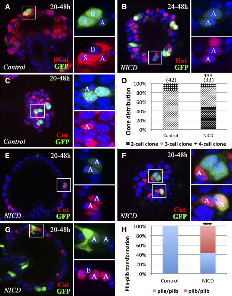
Figure 5.
Notch signaling is sufficient for pIIb fate. Flip-out clones are marked by GFP expression (green in A–C and E–G). (A) A representative control clone induced at 20 hr APF shows weak Notch activity in the AC and strong Notch activity in the SC. βGal expression (red) from Su(H)GBE-LacZ reporter is used to mark Notch activity. (B) Representative NICD-overexpressing clones induced at 24 hr and examined at 48 hr APF. The square area is magnified in the right two panels and shows a clone with two AC-like cells with faint GFP (green) and Hnt (red) expression. (C) Representative control clones induced at 20 hr and examined at 48 hr APF. The three-cell clone in the square area shows the AC with Cut expression (red). The other two clones have the same composition. (D) Quantification of clone distribution according to clone size. Clones were induced at 20 hr APF and examined at 48 hr APF. (E–G) Representative NICD-overexpressing clones induced at 20 hr and examined at 48 hr APF. (E) A two-cell clone (faint GFP) is composed of two ACs (Cut+). (F) A three-cell clone (upper panel) contains one AC (Cut+); a two-cell clone (one with faint GFP and one with strong GFP; lower panel) is composed of two ACs (Cut+). (G) A three-cell clone is composed of one EC (strong GFP and distinct cellular morphology) and two ACs (Cut+). (H) Quantification of clone distribution according to clone composition. pIIa/pIIb: clones containing one pIIa and pIIb cell during division. pIIb/pIIb: clones containing two pIIb cells during division, which gives rise to two ACs at 48 hr APF. Fisher’s exact test was used (***P < 0.001). AC, apical cell; APF, after puparium formation; βGal, β-galactosidase; EC, epithelial cell; GFP, green fluorescent protein; Hnt, Hindsight; NICD, Notch intracellular domain; SC, secretory cell; Su(H), Suppressor of Hairless.

