Figure 8.
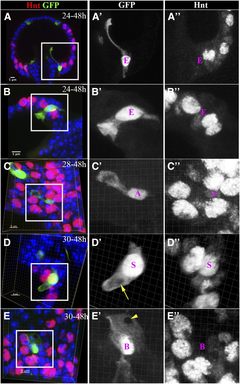
Cellular morphology of spermathecal lineage cells. (A and B) Single-EC clones induced at 24 hr and examined at 48 hr APF. (A) The EC localized at the junction of spermathecal lumen, and the duct has a long cytoplasmic protrusion in line with the introvert. (B) The EC localized in the middle region of the spermathecal head has an inverted umbrella-shaped apical membrane protruding into the lumen. The cytoplasm is marked by GFP [green in (A and B) and white in (A’–B’)]. (C) A single-AC clone shows the AC with an apical cytoplasmic bulge (C’). The AC is recognizable by the faint Hnt expression (white in C”). (D) A single-SC clone shows a finger-like apical protrusion with a hole in the middle (yellow arrow in D’). The SC is marked by strong Hnt expression (white in D”). (E) A single-BC clone shows a mesh-like apical membrane with a hole in the apical tip (arrowhead in E’). The BC is recognizable by the absence of Hnt expression (white in E”). The images in (B–E) are generated from three-dimensional volume rendering. AC, apical cell; BC, basal cell; EC, epithelial cell; GFP, green fluorescent protein; Hnt, Hindsight; SC, secretory cell.

