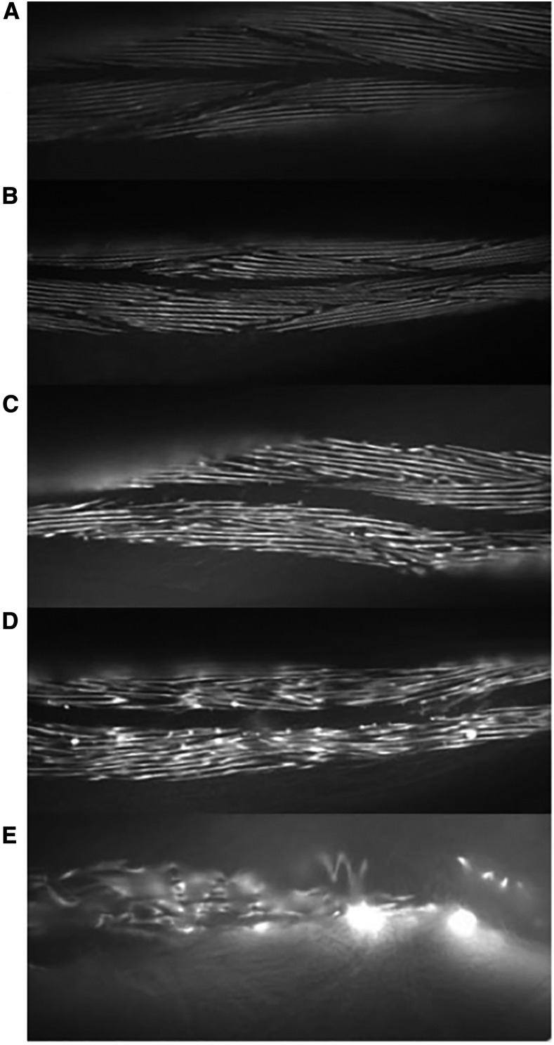
Figure 4.
Examples illustrating the five grades in the muscle disorganization scoring scale. The myosin::gfp fusion protein is localized to the thick filaments and so distribution of the fluorescence reports on the regularity in the arrangement of the sarcomeres. Images captured by fluorescence microscopy. (A) Typical structure of a grade 1 muscle score; myosin filaments are linear and well organized. (B) Typical structure of a grade 2 muscle score; myosin filaments are starting to show more bends but the pattern is still well organized. (C) Typical structure of a grade 3 muscle score; myosin filaments are more fragmented and there are apparently overlapping filaments. (D) Typical structure of a grade 4 muscle score; myosin filaments are further fragmented and the regularity of pattern is no longer clear. (E) Typical example of grade 5 muscle score; the pattern of myosin filaments is severely disorganized. Figure compiled by Matt Pipe.

