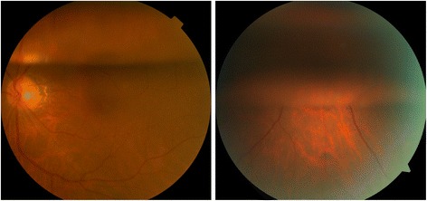
Fig. 2.

Typical fundus images of patients included in the study on postoperative Day 1. Group A (left): the air bubble remained at the top one-third of the vitreous cavity, with transparency of the visual axis. Group B (right): the air bubble remained at the top three-fifths of the vitreous cavity, without transparency of the visual axis
