Fig. 4.

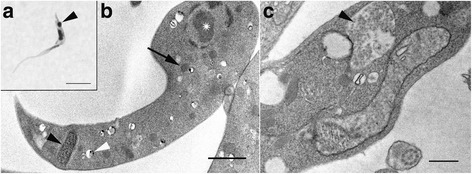
Trypomastigote of Trypanosoma sp. AAT. a Light micrograph exhibiting trypomastigote recognisable by posterior kinetoplast (arrowhead). b Transmission electron microscopy of trypomastigote exhibiting the kinetoplast (black arrowhead), nucleus (asterisk), acidocalcisomes (white arrowhead), and glycosomes (black arrow). c Reservosome in Trypanosoma sp. AAT (arrow). Scale-bars: a, 5 μm; b, c, 1 μm
