Fig. (1).

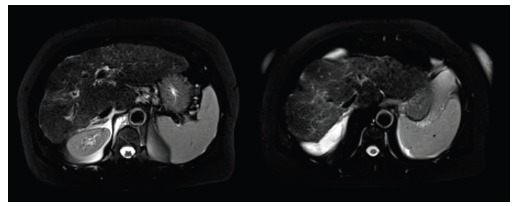
MRI, axial T2-weighted fat-suppressed images. Features of cirrhosis are visible: nodular surface, enlargement of caudal lobe, hyperintense bands within the liver as result of proliferation of fibrotic tissue, peritoneal effusion.

MRI, axial T2-weighted fat-suppressed images. Features of cirrhosis are visible: nodular surface, enlargement of caudal lobe, hyperintense bands within the liver as result of proliferation of fibrotic tissue, peritoneal effusion.