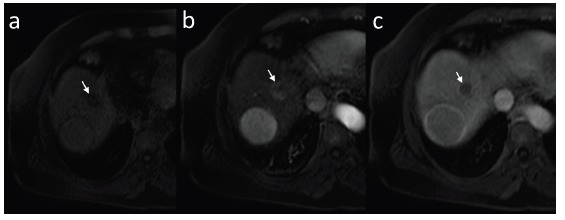
Fig. (15).

Small (arrow) and large HCC foci in T1-weighted fast-suppressed axial images: pre-contrast (a), in hepatic arterial phase (b) and equilibrium phase (c). Both lesions show enhancement in arterial phase and wash-out in equilibrium phase. Large lesion presents with enhancing pseudo-capsule in equilibrium phase.
